
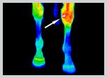
 Horse presented with non-specific intermittent lameness. Nerve blocks and radiographs could not determine the source. DITI detected inflammation on the medial left knee. Subsequent radiograph confirmed a small chip fracture at the exact location as indicated on the thermogram.
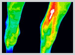
Horse presented with inflammation on left hock. EquiSpectrum DITI showed significant thermal findings on the anterior left hock consistent with a low grade infection.
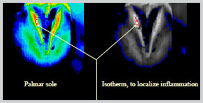
Horse presented with difficult to diagnose front hoof lameness. EquiSpectrum DITI detected a small puncture would that had become infected. Isotherm images were used to localize the inflammation.
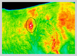
A standardbred trotter was reported by the trainer to not be “stretching out” or “moving well”. A hind limb or back issue was suspected to be the source of the discomfort. DITI showed no abnormalities in the limbs, but a well defined area of inflammation was seen over the right kidney region. Further testing confirmed the horse was suffering from renal pain and was treated successfully.
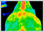
Horse presented with inflammation over four thoracic vertebrae in the withers and was scanned for suspected sacral issues. The injury was attributed to a damaged saddle tree.